English
The alveoli, along with the respiratory bronchioles, constitute what we call the respiratory zone. This is where gases are exchanged between the air in the alveoli and the blood in the surrounding capillaries (Fig. 5). The barrier between the air and blood is therefore very thin here. The epithelium in the alveoli is a single-layer of squamous epithelial cells (called type 1 cells). Thus, the barrier between blood and air is histologically as thin as it can be.
In addition to the capillaries, the alveoli are surrounded by elastic fibers. The elastic fibres clamp alveolar sacs together. This gives the lung tissue elastic properties that can stretch (when breathing in) and contract back again (when breathing out), the latter is a passive process not requiring energy.
The alveoli are organized into ”clusters” of air filled bladders. Pores between neighbouring alveoli allow air to diffuse from one alveolus to another. The gases in the lungs therefore diffuse across the alveoli so the concentrations of the various gases are relatively similar across all alveoli in the respiratory zone.
In the alveoli, in addition to the type-I-cells that form the alveoli wall, other cell types are present (Fig. 6). The macrophages belong to the immune system and ‘eat’ particles, dust and invading microorganisms that may enter the alveoli. Type II cells are cells that secrete surfactant (see below).
Inside the alveoli there is a thin film of water. The alveoli are not dry. This moisture is essential to maintain the diffusion of gases through the cell membrane, including the alveoli wall. Gases do not diffuse through dry membranes.
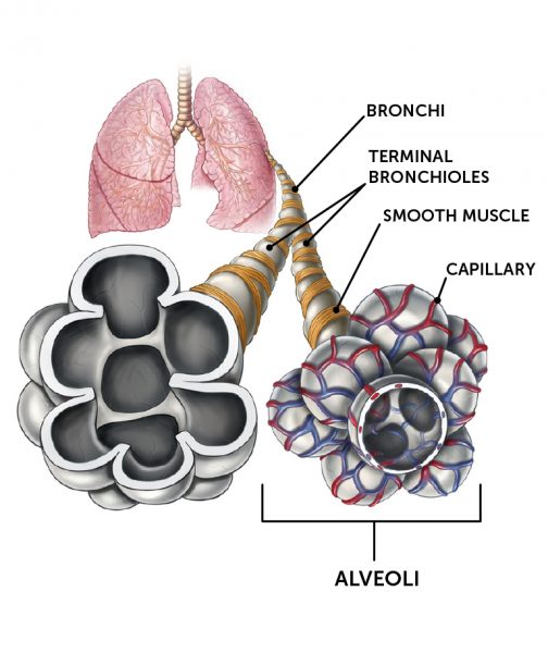
Fig. 5
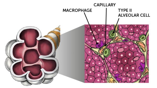
Fig. 6